Hip And Leg Bone Diagram - Bones Of The Leg And Foot Interactive Anatomy Guide - Bones give your body structure and enable you to move, but what else is your skeletal system responsible for?.
Hip And Leg Bone Diagram - Bones Of The Leg And Foot Interactive Anatomy Guide - Bones give your body structure and enable you to move, but what else is your skeletal system responsible for?.. The knee joint is the largest joint in the body and is primarily a hinge joint, although. Quizzes on human skeletal system anatomy, bone anatomy, and bone. Download 2,751 bone diagram stock illustrations, vectors & clipart for free or amazingly low rates! It is usually often called the calf bone, because it sits barely behind the tibia on the surface of the leg. Label bone diagram leg bone hip bone anatomy
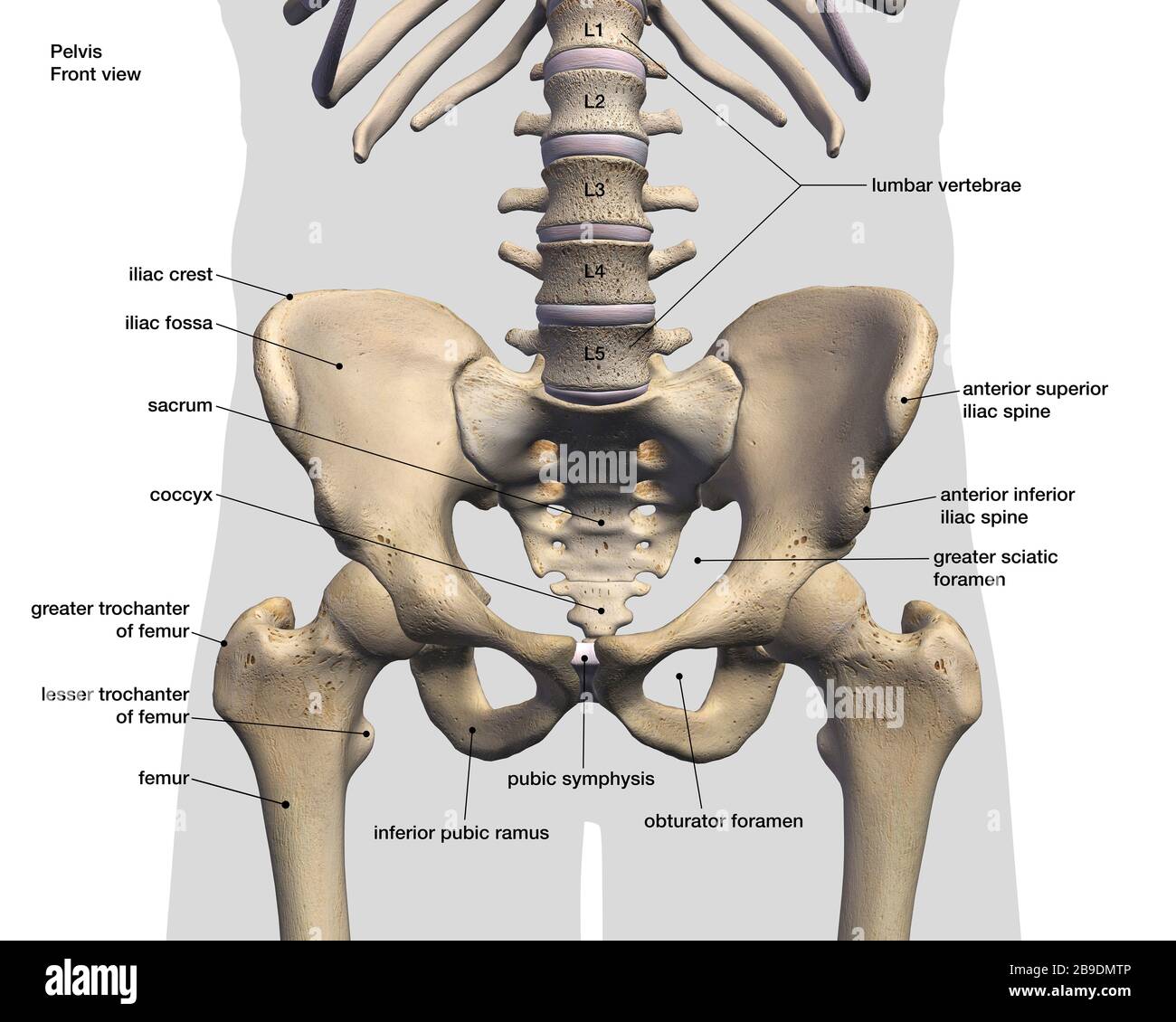
Feet human anatomy bones tendons ligaments and more. A diagram of the human skeleton. In this video you will learn the anatomy of the lower appendicular skeleton. This human anatomy diagram with labels depicts and explains the details and or parts of the hip bone structure. The foot bones shown in this diagram are the talus, navicular, cuneiform, cuboid, metatarsals and calcaneus.
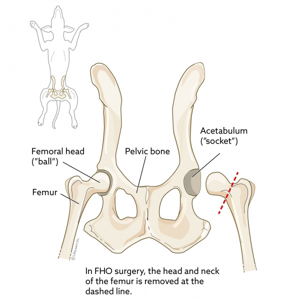
Femoral Head Ostectomy Fho In Dogs Vca Animal Hospital from vcahospitals.com Feet human anatomy bones tendons ligaments and more. The hip joint gives the leg an incredible range of motion while still providing support to the body's weight. It joins the lower limb to the pelvic girdle. When you stand or walk, all the weight of your upper body rests on them. Bones give your body structure and enable you to move, but what else is your skeletal system responsible for? Learn how to to left from and right and the meaning behind the names of the. The ilium bone forms the superior portion of the os coxa, the ischium bone the lower posterior portion, and the pubic bone (pubis) the lower anterior portion. The hip joint is made up of two bones:
The knee joint is the largest joint in the body and is primarily a hinge joint, although.
The knee is a strong but flexible hinge joint that uses muscles and. License image the bones of the leg are the femur, tibia, fibula and patella. Quizzes on human skeletal system anatomy, bone anatomy, and bone. Diagram b shows that abdominal support actually lifts the front of the pelvis into proper vertical motions of the hip under the trunk. Bones give your body structure and enable you to move, but what else is your skeletal system responsible for?. Basic bone diagram enthusiast wiring diagrams. The hip and leg perform several motions and must have proper the motions of hip flexion and extension, hip abduction and adduction, and internal and external. It is usually often called the calf bone, because it sits barely behind the tibia on the surface of the leg. Leg bones diagram femur manual e books. Hip anatomy, function and common problems. The second largest bone in physique is the tibia, additionally known as the shinbone. These same nerves innervate the knee, which explains why pain can be referred to the knee from the hip and vice versa. A diagram of the human skeleton.
Bones of the hip joint. Bones of leg and foot. Each leg is made up of four bones. Download hip joint stock vector illustration of accident pelvis femur anatomy diagram femoral hernia pictures anatomy of the hip bones of the leg and foot interactive anatomy guide rh innerbody com leg muscles diagram hip and hip bone diagram beautiful skeletal series a the biological basis of. Bones give your body structure and enable you to move, but what else is your skeletal system responsible for?
Diagram Human Body Bones Diagram Full Version Hd Quality Bones Diagram Mediagrame Tanzolab It from www.medicalartlibrary.com Disposition of rotator cuff muscles diagram. Each leg is made up of four bones. Download this free vector about diagram showing the hip bone treatment, and discover more than 15 million professional graphic resources on freepik. Download 2,751 bone diagram stock illustrations, vectors & clipart for free or amazingly low rates! The head of your femur fits into your hip socket and the bottom end connects to your knee. Labeled skeleton diagram best of pelvic bones simple bone diagram. It joins the lower limb to the pelvic girdle. Quizzes on human skeletal system anatomy, bone anatomy, and bone.
The knee is a strong but flexible hinge joint that uses muscles and.
At the distal end of the femur, two rounded condyles meet the tibia and fibula bones of the lower leg to form the knee joint. Your leg bones are the longest and strongest bones in your body. The bones of the leg are the femur, tibia, fibula and patella. In this video you will learn the anatomy of the lower appendicular skeleton. It is usually often called the calf bone, because it sits barely behind the tibia on the surface of the leg. Hip anatomy, function and common problems. Bones of the hip joint. The foot bones shown in this diagram are the talus, navicular, cuneiform, cuboid, metatarsals and calcaneus. Front view of the hip joint bones. Basic bone diagram enthusiast wiring diagrams. Normally, a smooth cushion of shiny white hyaline (or articular) gluteus medius and minimus are the main abductors of the hip —that is, they move the leg away from the midline of the body (using the spine as a midline. Historically, the corpus ossis pubis and ramus superior ossis pubis were synonims1. The foot bones shown in this diagram are the talus, navicular, cuneiform, cuboid, metatarsals and calcaneus.
Bones of the hip joint. Bones of leg and foot. Your leg bones are the longest and strongest bones in your body. Learn how to to left from and right and the meaning behind the names of the. Feet human anatomy bones tendons ligaments and more.
Labeled 3d Medical Illustration Of Male Pelvis Hip And Leg Bones On White Background Stock Photo Alamy from c8.alamy.com The knee joint is the largest joint in the body and is primarily a hinge joint, although some sliding and rotation occur. Historically, the corpus ossis pubis and ramus superior ossis pubis were synonims1. Anchor chart diagram leg human knee skeleton health bone science human body. Find the perfect bone diagram stock illustrations from getty images. Leg bones diagram diagram schematic ideas. The foot bones shown in this diagram are the talus, navicular, cuneiform, cuboid, metatarsals and calcaneus. Front view of the hip joint bones. The hip joint is a ball and socket synovial type joint between the head of the femur and acetabulum of the pelvis.
This human anatomy diagram with labels depicts and explains the details and or parts of the hip bone structure.
Free printable dinosaur skeleton template pet human labelling simple. Learn how to to left from and right and the meaning behind the names of the. Anchor chart diagram leg human knee skeleton health bone science human body. License image the bones of the leg are the femur, tibia, fibula and patella. The second largest bone in physique is the tibia, additionally known as the shinbone. The hip joint gives the leg an incredible range of motion while still providing support to the body's weight. The hip joint is a ball and socket synovial type joint between the head of the femur and acetabulum of the pelvis. Labeled skeleton diagram best of pelvic bones simple bone diagram. Human anatomy diagrams and charts show internal organs, body systems, cells, conditions, sickness and symptoms information and/or tips to ensure one lives in good health. Leg bones diagram femur manual e books. Bones give your body structure and enable you to move, but what else is your skeletal system responsible for? The two bones beneath your knee that make up your shin are. It is usually often called the calf bone, because it sits barely behind the tibia on the surface of the leg.
Fibula and tibia, ankle and foot leg bone diagram. Fibula and tibia, ankle and foot.


0 Komentar